
Biotite
Biotite or black mica is a member of the mica group within the sheet silicates. Its general formula is K(Mg,Fe)3(AlSi3O10)(OH)2. Biotite is usually a solid solution of several minerals, mostly Fe-rich annite, KFe3(AlSi3O10)(OH)2 , and Mg-rich phlogopite, KMg3(AlSi3O10)(OH)2; the exact composition of a sample is variable and has to be determined through a thorough analysis.
Crystals are six-sided pseudorhombohedral (no right angle between vectors; vectors of different lengths) or eight-sided pseudohexagonal (meaning they have no hexagonal symmetry). In common language it means crystal faces are parallelograms or hexagons with sides of different lengths. Both types of crystals are made of several six-sided monoclinic unit cells. They have only one perfect cleavage and so break into sheets as shown on figure 1.
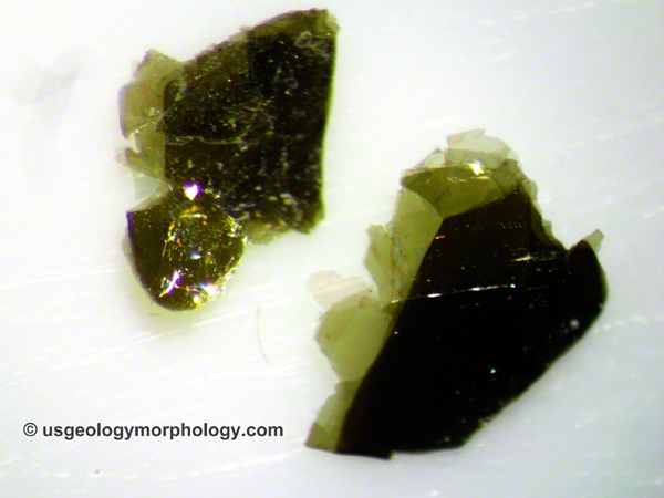
Figure 1. Biotite crystals. Biotite is commonly called black mica. Width of view: 4 mm.
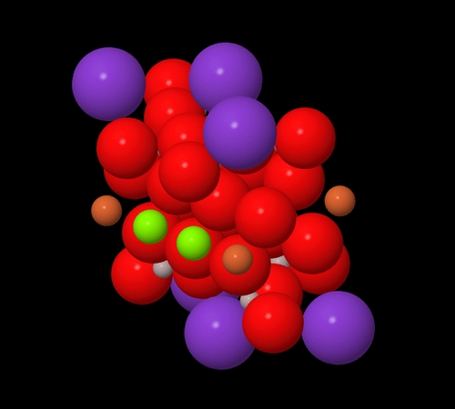
Figure 2. Atomic structure a biotite sample: atoms at actual size.
This cartoon shows one unit cell with its atoms at nearly actual size. At their actual size, some would be a little bigger and all would be in contact with each other (principle of closest packing: anions and cations, attracting each other, come as close as their respective radii allow). The difficulty to see all the atoms and the impossibility to represent their bonds explains why the ball-and-stick models of figures 3, 4 and 5 are preferred. Atom colors: light-grey-Si (silicon) or Al (aluminum); red-O (oxygen); tan-Fe (iron); green-Mg (magnesium); purple-K (potassium); white-H (hydrogen). Data: Brigatti et al. (1990). Software: Jmol.
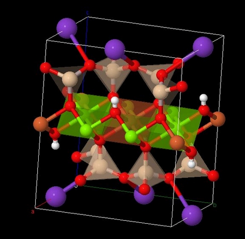
Figure 3. Crystal structure of a biotite unit cell.
Same sample as in fig. 2 in a ball-and-stick model. The white box is one unit cell (monoclinic). Atom colors: light-grey-Si (silicon) or Al (aluminum); red-O (oxygen); tan-Fe (iron); green-Mg (magnesium); purple-K (potassium); white-H (hydrogen). Polyhedra have the same color as the atoms around which they are organized. Note the silicon tetrahedra (one Si for four O, SiO4-). Al, which has a similar radius as Si, substitutes for Si in some tetrahedra. Each O atom has one electron left to bond with another Si (so Si/Al tetrahedra share oxygen atoms)or one of the following: Mg, Fe, K, H. See more about bonds and polyhedra in fig. 5. Data: Brigatti et al. (1990). Software: Jmol.
The Jmol application is written in Javascript. You may be asked to authorize its download and may have to add the complete url of this page to your exception list in the Java console. If it still doesn't work, restart your browser. See more about this program at the bottom of the page.
Use your mouse to move the figure; right-click for additional commands. Mouse over atoms to display their names. Same atom colors as in fig. 3.
Reload the whole page to reset the original view if necessary.
Figure 4. Crystal structure of a biotite sample: interactive view.
Same sample as in fig. 2 with two unit cells shown. Same colors, except the unit cell (blue box). Data: Brigatti et al. (1990). Software: Jmol.
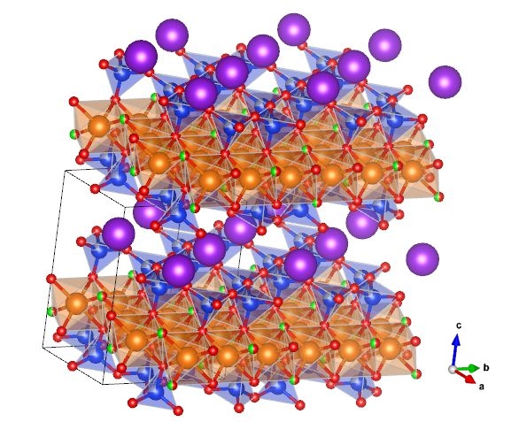
Figure 5. Atomic structure of a phlogopite (biotite) sample containing fluorine.
Formula: KMg3(AlSi3O10)(F,OH)2 . Atom colors: blue-Si (silicon); light-grey-Al (aluminum); red-O (oxygen) or OH- (hydroxide ion); tan-Mg (magnesium); purple-K (potassium); green-F (fluorine). Bicolor balls show alternative atoms. Unit cell in black. Phlogopite, as mentioned above, is an end-member of biotite; it has magnesium (Mg) but no iron (Fe). The cartoon shows two layers of silicon/aluminum tetrahedra (4 sides, light blue) "sandwiching" a layer of magnesium octahedra (8 sides, light tan). These layers form the three basic sheets of the phlogopite crystals; using the first letters of tetrahedron and octahedron, this structure is called "T-O-T". Notice that in each tetrahedron, three oxygen (out of four) link to other tetrahedra, forming tetrahedral planes: that's the distinctive feature of sheet silicates (or phyllosilicates from the Greek word phyllos - sheet or leaf -). In this sample, fluorine (F) atoms substitute for hydroxide ions (OH-). K+ cations are bonded to oxygen and link T-O-T units together; the weakness of their bonds explains why it's generally here that biotite crystals break, i.e. along the (0,0,1) face. Data: Hendricks et al. (1939). Software: VESTA (Momma et al., 2011).


Click on each individual image for full-size zoomable image
Figure 6. Thin section of biotite in Morton gneiss, Minnesota River Valley.
This tonalite gneiss, mostly composed of quartz, plagioclase and biotite is one of the oldest rocks in the U.S. (~3.5 Ga). In plane-polarized light (left), biotite is brown-green to brown, depending on the orientation of the crystals; quartz and plagioclase are colorless. In crossed polarizers (right), biotite is brown to green-brown; the grain at lower right is near extinction and shows typical biotite mottled extinction pattern. Field of view: 2.3 mm. Bt-biotite; Pl-plagioclase feldspar; Qtz-quartz
More microphotographs of Morton gneiss here.
References:
Brigatti, M. F., and Davoli, P., 1990, Crystal structure refinement of 1M plutonic biotites, American Mineralogist, v. 75, p. 305-313.
Hendricks, S. B., and Jefferson, M. E., 1939, Polymorphism of the micas with optical measurements Note: Biotite group, American Mineralogist, v. 24, p. 729-771.
Momma, K., and Izumi, F., 2011, VESTA 3 for three-dimensional visualization of crystal, volumetric and morphology data, J. Appl. Crystallogr., v. 44, p. 1272-1276.
Jmol is a free, open-source Java 3D viewer for chemical structures, offered under the GNU Lesser General Public License. More details at http://wiki.jmol.org/.